
Ventilation Perfusion Scan
So you hear the doctors talking about the patient going down to interventional radiology (IR) for a "VQ scan." As a nurse, you should know what the purpose of this scan is. So what is this and why does it matter?
What Happens During a VQ Scan?
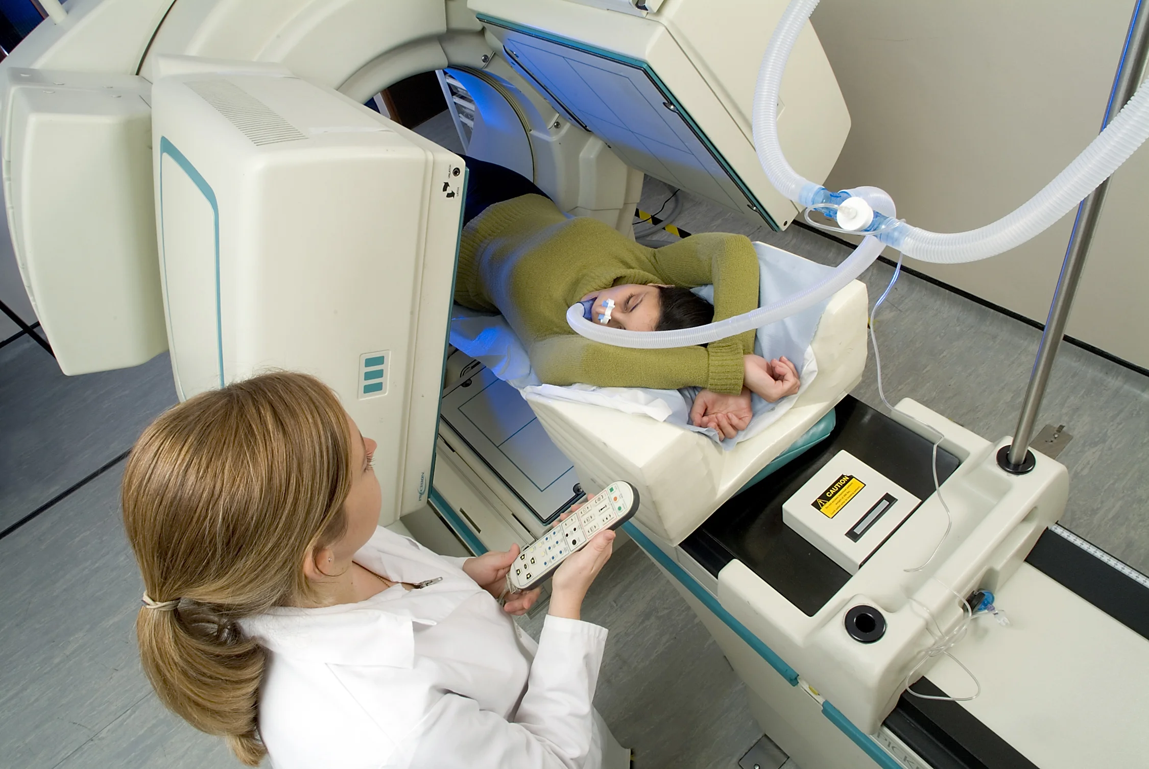
A VQ scan, more properly known as a ventilation perfusion scan, is used to examine two different elements within the lungs: air flow and blood flow.
The test is performed in two parts to test these. The first part of the test involves having the patient inhale radioactive material and then taking pictures of the lungs as it is inhaled. In the second part, the patient has radioactive material injected into an intravenous line (IV) and then pictures are taken to see the blood flow in the lungs. The two sets of images are then compared with each other.
Why?
The main reason to have this test performed is to see if a patient has pulmonary blood flow issues. The most common reason I have seen is to check if there is a pulmonary emboli (a blood clot in the lungs). This can be fatal as it blocks off essential blood flow to the lungs. When you think of a heart attack, you think of something blocking vessels that are surrounding the heart causing the infarction. With a pulmonary embolism, it is the same idea but in the lungs. You have a clot that is blocking that blood flow. On imaging during the VQ scan, the interventional radiologist can see if there is compromised blood flow which then allows us to treat this condition.
Imaging Examples
Here are a couple examples of imaging that might be seen on an abnormal VQ scan:

"Perfusion scan from a patient with severe chronic thromboembolic pulmonary hypertension. The right lung has nearly no blood flow. The left lung has multiple wedge shaped blood flow defects." -Pulmonary Hypertension RN

"The ventilation scan is from the same patient. The left lung has preserved ventilation. These findings are classic for CTEPH." -Pulmonary Hypertension RN
Other relevant articles include:
References